
 Home
Home Health
 Diet & Nutrition
 Living Well
 More
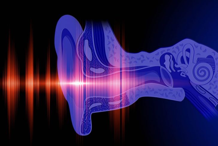
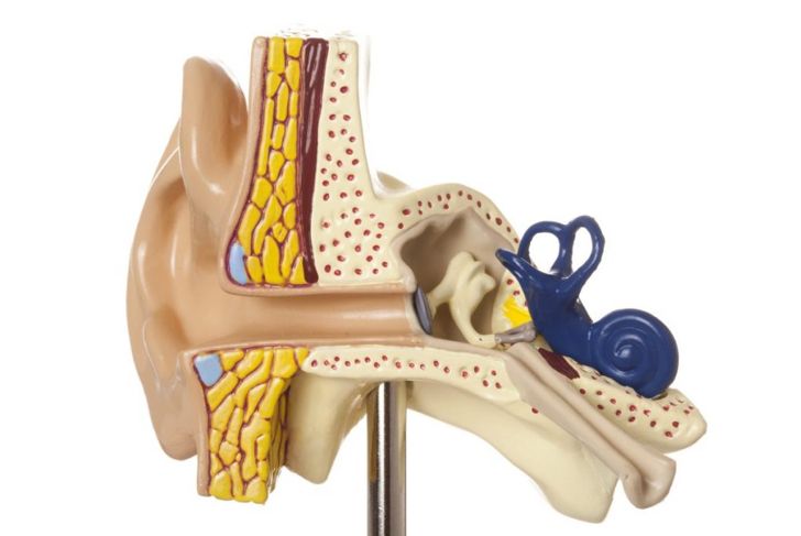
MoreA cholesteatoma is a noncancerous skin growth made up of squamous epithelium cells. It develops in the middle of the ear, behind the eardrum. The cholesteatoma is a skin-lined cyst composed of trapped living and dead skin cells and other debris. The dead skin cells accumulate in and around the living tissue inside the cyst. As the cyst grows, the structures of the middle ear are damaged. If this condition is not treated, it eventually results in hearing loss, vertigo, and even facial muscle paralysis.
Initial Symptoms
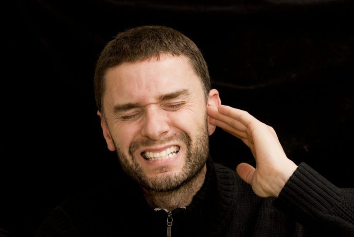
At first, symptoms of cholesteatomas are usually mild. Hearing loss and ear pain are the most common initial symptoms. Cholesteatomas grow in only one ear, so symptoms are limited to that ear and the corresponding side of the face. Mild drainage or increased ear wax secretions may develop as well. A doctor examining the ear during the early stages may observe skin cell deposits or masses of blood vessels.
Mechanism of Damage
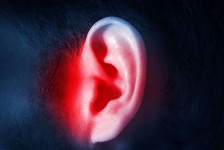
Cholesteatomas damage the middle and inner ear by eroding bone, cartilage, and tissue as the cyst expands. This process generally occurs over years. Hearing loss happens first when damage occurs to the delicate ossicles in the middle ear. Vertigo is a common symptom once the cholesteatoma reaches the inner ear. The mix of dead and living skin cells, blood vessels, and other tissues in the cyst attract bacteria or fungus that create a chronic infection. A cholesteatoma can be fatal if it grows into the cranium and the infection causes an abscess in brain tissue.
Causes
Cholesteatomas can form after injury to the eardrum, a chronic middle ear infection, or chronic sinus pressure. Continuous pressure in the sinuses weakens the eardrum and leads to small pockets below it that fill with trapped skin cells and debris. Eustachian tubes are small openings connecting the middle ear to the nose. These tubes equalize pressure in the ears, so when the tubes are blocked or not functioning properly, the pressure damages the eardrum.

Advanced Symptoms
A growing cholesteatoma eventually leads to amber, white, or blood-tinged fluids draining from the ear. The fluid has a foul smell and thick consistency. Affected people feel pressure, and constant, aching pain develops in and behind the ear. Pain often spreads into the sinuses and dizziness, irreversible hearing loss, and facial muscle paralysis develops as the cyst grows and spreads.
Complications
Without intervention, cholesteatomas continue growing and inflicting more damage. A large cholesteatoma that has grown into surrounding tissue or formed a complex sac throughout the ear may cause permanent hearing loss. Vertigo and poor balance when standing or walking may also be permanent if extensive damage in the middle ear or destruction of sensitive nerves in and around the ear occurs before treatment.

Risk Factors and Prevention
Recurring middle ear or sinus infections are the most common risk factors for developing a cholesteatoma, and a family history of middle ear infections can also increase the risk. Smoking or living in a home with second-hand smoke greatly increases the likelihood of these precursory conditions. Congenital cholesteatomas begin developing at birth. This is rare but can be managed by monitoring growth and intermittent removal to prevent cysts.

Evaluation
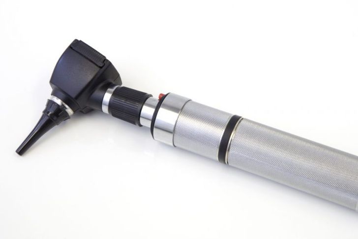
Numerous tests can help medical professionals diagnose and evaluate cholesteatomas. The initial exam includes a visual assessment of the eardrum using an otoscope and tuning fork test to determine conductive hearing loss. An audiogram measures hearing loss while a tympanogram shows perforation of the eardrum. An MRI scan shows the soft tissue in the middle ear and helps detect recurrent or residual cholesteatoma tissue. A CT scan of the temporal bone can indicate bone erosion and other damage.
Surgery
Cholesteatomas almost require surgical removal. The cholesteatoma is removed from the eardrum, the middle ear and the space behind the eardrum. Multiple surgeries are common because it is difficult to remove all of the skin cells and other tissue in the cyst at once. Additional surgeries check for recurrence — sometimes the growths expand between surgeries, or existing cysts hide additional growths upon examination.
Surgery Complications
The ear and surrounding areas contain small, fragile bones, nerves, and delicate tissue. Surgery is challenging, and sometimes surgeons create extra holes unintentionally. These can lead to canal fistulae between the ear canal and other areas of the ear, or between cavities in the ear and cranial vault. The holes cannot heal correctly on their own and frequently become infected so patients may require antibiotics and additional surgeries. Surgeons must take care to avoid damaging nerves during surgery because these cannot be repaired. Sometimes, large or complex cholesteatomas make nerve damage unavoidable.

Prognosis
The prognosis after treating and removing a cholesteatoma is good if medical practitioners catch and correct the issue before severe complications occur. The hearing usually improves immediately and remains stable after surgery. Surgeons can at least partially repair physical damage to the small bones in the ear, the ossicles, and the tympanic membrane forming the eardrum. Patients can take antibiotics orally or as ear drops before and after surgery to limit infection. Lifelong monitoring is necessary to catch the recurring growth of cholesteatomas, which is common.