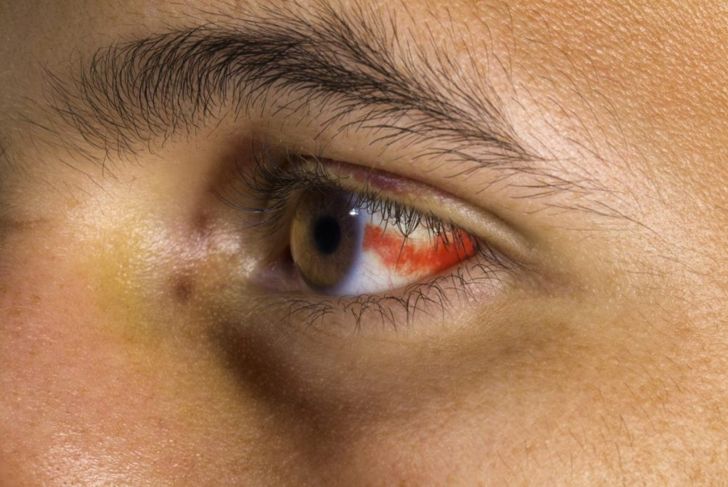
A subconjunctival hemorrhage occurs when a tiny blood vessel breaks just underneath the conjunctiva. Sub means under, and hemorrhage means bleeding. The event can have various causes that are harmless and require no treatment and others that warrant a visit to the doctor. Physical damage to the surface of the eye often involves the conjunctiva. Causes include thermal or chemical burns and blunt or penetrating trauma. While the extent injury may be limited to the conjunctiva, subconjunctival hemorrhage may be a result of a more serious, underlying problem. Careful evaluation, initial management, and triage of conjunctival injuries are essential to promote healing.
How often does it happen?
A subconjunctival hemorrhage may look scary but is a common condition. There are more than two hundred thousand cases per year. Treatment can help but cannot cure it. It’s easily identifiable and requires no lab tests or imaging. The whole white part of the eye may turn red, or just part of it. Blood in the eye can happen at all ages. Often the causes in younger people are different than those for older adults.
Causes of Subconjunctival Hemorrhage
Subconjunctival hemorrhages have various underlying causes including significant blunt trauma to the eye, minor trauma or complications from contact lens use, history of elevated venous pressure (coughing or vomiting), hypertension, history of diabetes mellitus, or bleeding disorders. It can also be spontaneous. The condition is common in the elderly due to hypertension. They are also common in newborns and may persist for several weeks. However, in infants with no verifiable history of trauma, healthcare practitioners may suspect child abuse.
Conjunctiva
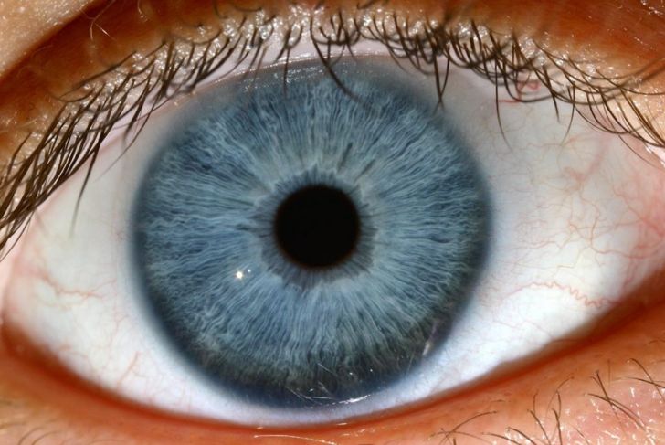
The conjunctiva is divided into the palpebral or tarsal conjunctiva, bulbar or ocular conjunctiva, and fornix conjunctiva. It is made up of special types of skin cells called stratified columnar epithelium and unkeratinized, stratified squamous epithelium with goblet cells. The palpebral or tarsal conjunctiva lines the eyelid. The bulbar or ocular conjunctiva covers the eyeball. The fornix conjunctiva is a loose and flexible junction between the bulbar and palpebral conjunctivas that allows free movement of the lids and eyeball.
Blood Vessels of the Eye
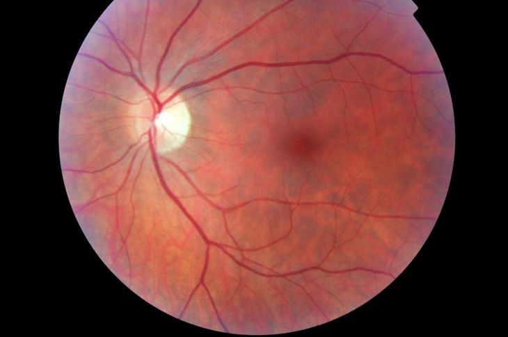
Small, branch arteries of the ophthalmic artery deliver blood to the eye. The ophthalmic artery branches from the internal carotid artery. The smaller arteries include the central retinal artery, the short and long posterior ciliary arteries, and the anterior ciliary arteries. The anterior ciliary arteries, smaller vessels from the anterior choroid, and posterior ciliary arteries supply the iris and ciliary body in the vascular tunic. They travel by different paths to eventually join the major arterial circle of the iris.
Signs
When any of the blood vessels rupture due to the aforementioned causes, this releases blood into the surrounding anatomical environment. It appears as a bright red patch in the white of the eye. The blood is below the conjunctiva, hence the name subconjunctival hemorrhage, and will not leak. The blood will stay there for two to three weeks depending on the patient due to the conjunctiva’s slow absorption of blood.
Symptoms
Often, no symptoms accompany a subconjunctival hemorrhage, but some may feel a foreign body sensation or a scratchy feeling on the surface in the eye. Patients who take blood thinners should be evaluated for overmedication if subconjunctival hemorrhages are recurrent or occur with other signs of a bleeding disorder, such as bleeding gums. Elderly patients may have elevated blood pressure. The condition does not affect vision, cause discharge from the eye, or cause pain.
Complications
Patients with traumatic subconjunctival hemorrhage may have ruptures in the integrity of the eyeball or blood in the anterior chamber of the eye. Scleral laceration or rupture may elevate subconjunctival hemorrhages and hide the laceration. Other possible eye injuries to consider are lacerations, abrasions, or foreign bodies. Individuals can treat small lacerations and abrasions with antibiotics. Large lacerations require further medical inspection and treatment. A doctor will remove foreign bodies with a cotton-tipped applicator and topical anesthetic.
Action
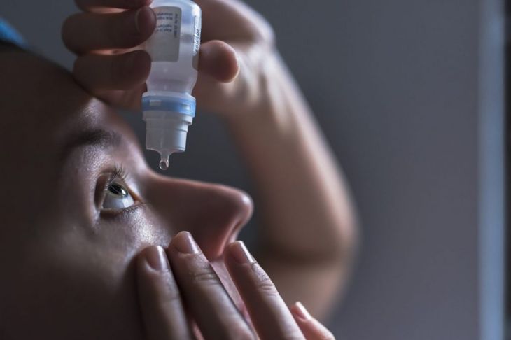
Most of the time, individuals do not notice subconjunctival hemorrhages until someone else points them out. Blood in the eye that results from trauma, or cases when trauma cannot be ruled out, may warrant urgent ophthalmology consultation. If the cause is a spontaneous, non-traumatic incident, most of the time it will resolve on its own in two to three weeks, requiring no further medication or treatment. If the hemorrhage does not resolve, the individual should seek professional medical assistance.
Questions to Consider
A doctor will collect a concise history of events before the patient noticed the subconjunctival hemorrhage. They will also examine the affected eye and check the patient’s blood pressure. If trauma is the cause, he or she will examine the eye using a slit lamp. While seeing the physician, patients may want to inquire
Is there any damage to the eye?
How will this affect my vision?
What are the possible causes of blood in the eye?
What can I do to prevent this from happening again?
Outcomes
Often, there is no major cause for concern if a subconjunctival hemorrhage spontaneously appears. Patients like these typically recover with no loss of vision. However, if one’s medical history includes diabetes or hypertension, blood thinning medications, past subconjunctival hemorrhages, traumatic injury, or other concurrent conditions, it is advisable to see a doctor. In such cases, visual outcomes depend on the severity of the injury.

 Home
Home Health
 Diet & Nutrition
 Living Well
 More
More
