
 Home
Home Health
 Diet & Nutrition
 Living Well
 More
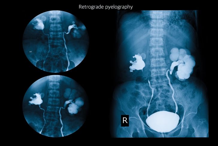
MorePyelography is the technique of taking photographs of the kidneys, ureter, and bladder. Your Ureter carries urine from a kidney to the bladder. These photographs are done using X-rays. To perform a pyelography, a patient must be injected with an opaque solution or a radiopaque dye. A radiopaque dye becomes visible in both an X-ray and fluoroscopy. The procedure is often called an intravenous pyelogram. It is not as scary as it sounds. In Fact, an intravenous pyelogram also called an IVP, may allow your doctor to treat your condition with medication, and avoid surgery.
When Does a Doctor Order a Pyelogram?
Most often a doctor will order a pyelogram if there is suspected obstruction to the flow of urine. Usually, this means the doctor suspects a blockage due to a kidney stone. Patients with problems related to the kidneys might also need to undergo this test as it provides information about the functioning of the kidneys. If your doctor suspects you have any problem with your kidney, or urinary tract this is most often the first test ordered. Your doctor may even order a CT scan done at the same time, as a CT scan also uses contrast dye.
Why might I need an Intravenous Pyelogram?
A pyelogram can show your doctor the size, general state, and structure of your kidneys, ureters, and bladder. If you have or your doctor suspects you have any of the following conditions, you may need this test performed:
- Kidney disease
- Ureter or bladder stones
- Enlarged prostate
- Trauma or injury to the urinary tract
- Tumors
- Pain in your side or kidney area

What Are The Risks of a Pyelogram?
There are two types of dyes used in an intravenous pyelogram, ionic and nonionic. Both types will contain some iodine, but each is different in two key areas. Both types of dye have an overall low rate of adverse effects, but the less expensive ionic dye is known for a greater chance of adverse reactions in patients. Most adverse reactions are minor, with flushing, nausea, vomiting, and itching being some of the complications. A very small percentage of patients will experience serious adverse reactions including difficulty breathing, low blood pressure, loss of consciousness and swelling of the lips and tongue.

Who is at Most Risk When Undergoing a Pyelogram?
Anyone with diabetes, high blood pressure, the elderly, heart disease, or evidence of dehydration have a risk of developing kidney failure after the test. For those with these conditions, special precautions that must be discussed with your doctor before this test can be performed. Kidney function can be tested via a blood test for creatinine before the pyelogram is performed. Your doctor should review the results of this test before any pyelogram. People with diabetes taking certain medications will need to stop taking this medication before and a few days after the pyelogram. If you have any of these conditions be sure to discuss this with your doctor.

How do I Prepare For an Intravenous Pyelogram?
This procedure is usually performed on an outpatient basis. It is rather straightforward to prepare for this test. Your doctor may order you to take laxatives. In some cases, your doctor may also order enemas be performed to cleanse out any stool which may interfere with the test results. You will most likely be asked not eat anywhere from eight to twelve hours before the test is performed.
What Happens During the Intravenous Pyelogram Procedure?
Your test will be performed in an X-ray department at either an outpatient facility or at a hospital. A dye will be injected via an IV while you are laying down. Several X-rays will need to be taken, enough so that the dye shows up on the X-ray. The dye will make the kidneys, bladder, and ureters appear stark white on the X-ray.After the organs can be seen on the X-ray, your doctor may have you urinate so they can take one final X-ray. Your vitals will be monitored during the test, and a nurse will be on hand to ensure you are not experiencing pain or nausea. Should you experience pain or vomiting during the procedure, your doctor may have the nurse administer the appropriate medicines through your IV.

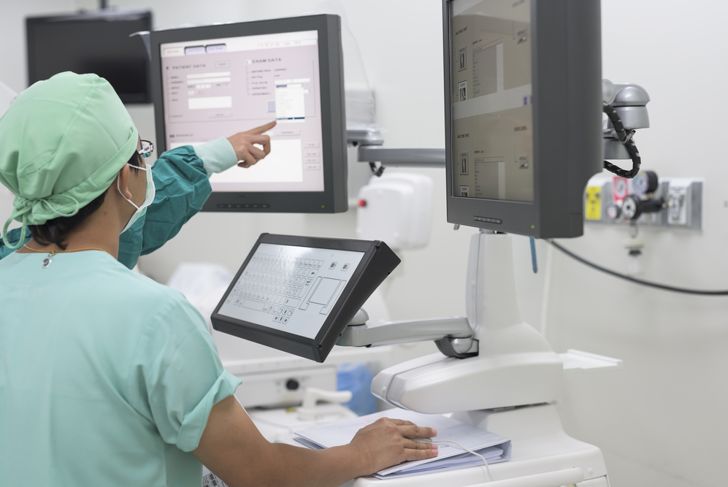
What does the equipment look like?
The equipment used for this test will typically consist of a radiographic table, one to two X-ray tubes and a monitor. A fluoroscopy will also be present, which includes an X-ray machine and a detector. The fluoroscopy is suspended over the gurney that you lie upon. The detector is used to produce the video or photos required for the test. A continuous X-ray beam is passed through any body parts being examined. This beam is then transmitted to a monitor that is often present in the exam room. The monitor is used by the technician to see the body part and its motion in detail.
What Happens After The Pyelogram Procedure?
After the X-rays are done, your doctor will review the results of the film. If the doctors were unable to find the cause of your problem, you might have to undergo further testing. If an obstruction was detected, you might need to be admitted to a hospital for surgery, such as in the case of a kidney stone that is too large to pass. You may be referred to a urologist if you haven’t already. In cases of an obstruction, surgery is not always needed; your doctor may order a lithotriptor procedure. This procedure uses sound waves to break the stone into smaller sizes that can be passed in the urine.

Who interprets the results and how do I get them?
A radiologist will analyze your test results. A radiologist is a physician specially trained to supervise and interpret radiology examinations. The radiologist will work with your primary care doctor or specialist, who will discuss any results with you. You may be required to do one or more follow-up examinations. If more exams are required your doctor will discuss with you the reasons why. Follow up exams are sometimes required if any abnormality needs further evaluation.

Pyelogram Aftercare
A pyelogram is a rather simple routine procedure. You will typically not need any special care after a pyelogram. You should, however, keep track of how much fluid you are consuming and how much urine you pass over the next 24 hours following the procedure. Drinking more fluids will help you to flush out the contrast dye from your body. You should contact your doctor if you experience any of the following after your pyelogram:
- Fever or chills
- Swelling, or bleeding from the IV site
- Blood in your urine
- Nausea, hives, itching, or sneezing