 Home
Home Health
 Diet & Nutrition
 Living Well
 More
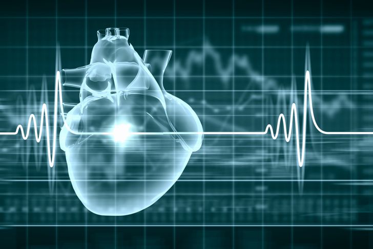
MoreAn echocardiogram is a test that helps doctors diagnose and monitor a variety of heart conditions by creating a moving picture of the heart. This picture helps medical professionals identify areas of the organ that are not working as they should, and enables early diagnosis and treatment to prevent complications.
What is an Echocardiogram?
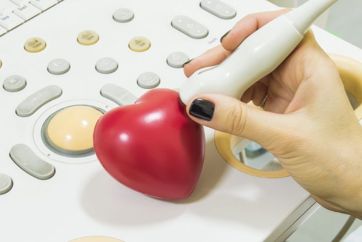
An echocardiogram is an ultrasound test that usesa transducerto send high-pitched sound wavesthrough the heart. As sound waves bounce off different locations within the organ, the device records the echo. The findings are then transformed into a moving picture that displays the patient’s heart activity onscreen. There are four types of echocardiograms: stress, Doppler, transesophageal, and transthoracic.
Stress and Doppler Echocardiograms
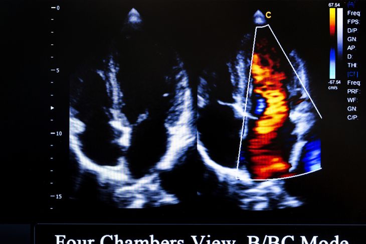
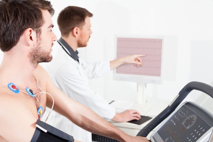
A stress echocardiogram might be administered during a stress test. It is performed both before and after the doctor applies the stressor, which is often exercise, usually on a treadmill or stationary bike, or medication that makes the heart beat more quickly. The echo measures blood flow along with other features of the heart. A Doppler echocardiogram uses a transducer to detect the movement of blood through the echoes of sound waves. It tests how blood flows through the chambers, valves, and vessels of the heart. The ultrasound also follows the speed and direction of the blood.
Transesophageal and Transthoracic Echocardiograms
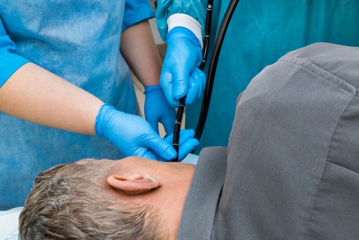
A transesophageal echocardiogram or TEE is a more invasive ultrasound because a thin probe is inserted down the throat and into the esophagus. Since the test is monitored from inside the body rather than the outside, it enables the doctor to capture clearer images of the upper chambers of the heart and their valves. Not only is it closer to the upper chambers of the heart, but the chest wall and lungs will not block any sound waves. Patients require a sedative for a TEE and medication to numb the throat. Transthoracic echocardiogram or TTE evaluates the heart size and wall thickness along with the blood flow and beats. This test is not invasive and does not require sedatives. Transthoracic is the most common type of echocardiogram.
Reasons for a Transthoracic Echocardiogram
A doctor may recommend a transthoracic echocardiogram to help diagnose, manage, or treat a heart condition. The test can:
- assess the risk of heart disease,
- monitor existing heart disease and the body’s response to treatment
- locate narrowed or blocked arteries, tumors, and blood clots,
- analyze symptoms such as chest pain, shortness of breath, or palpitations,
- discover which heart valves are not functioning,
- determine the heart walls that are not beating in synchronization, or
- assess tissue damage and overall activity after a heart attack.

Reasons for a Transesophageal Echocardiogram
A transesophageal echocardiogram can:
- monitor the heart during surgery,
- guide procedures during cardiac catheterization,
- check artificial heart valve functionality,
- inspect the left atrium, or upper left chamber of the heart, for blood clots,
- identify abnormal blood flow between the heart chambers or a cardiac shunt,
- test for endocarditis, an infection of the valves of the heart or the inner lining, or
- identify an aortic dissection, a tear in the aorta.

Reasons for Stress and Doppler Echocardiograms
Unlike TTE and TEE, a stress echocardiogram offers valuable information about heart function in response to exercise. A doctor may use one to monitor blood flow to the heart muscle — reduced blood flow can indicate ischemia, which is more noticeable after exercise. A Doppler echocardiogram can be used alongside a stress, transthoracic, or transesophageal echocardiogram to measure the speed at which blood travels through the heart.
Transthoracic Echocardiogram Procedure
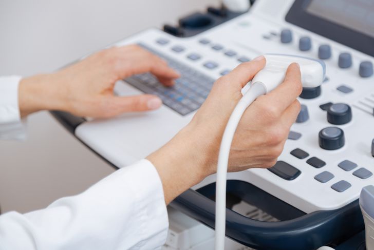
During the transthoracic echocardiogram, the patient removes their clothing from the waist up and puts on a gown. The medical practitioner will place three electrode pads on the chest and shoulders and have the patient lie down on their left side. He or she will use a cold, water-based gel and may have to apply pressure to the chest using the transducer to obtain clearer pictures. Noise may also come out of the device as it gathers information. Nothing about the test is painful, but it might be slightly uncomfortable. The test takes about 45 to 60 minutes.

Stress Echocardiogram Procedure
During a stress examination, the practitioner will perform two echocardiograms. The doctor will first complete a baseline test. After exercise, the technician will perform a second echocardiogram. If the doctor administers medication in place of exercise, the patient will lie down for the ultrasound, which requires electrodes to be affixed to the arms and legs and may include an intravenous line as well. The entire procedure takes about an hour.
Transesophageal Echocardiogram Procedure
A TEE is the most invasive variety. The patient will receive a throat-numbing spray, lozenge, or liquid and an IV medication to reduce saliva and stomach secretions. The IV may also have pain medicine or a sedative to relax the patient. Breathing rate, blood pressure, and heart rate will all be monitored during the test. The patient will lie on his left side and the doctor may ask him to swallow as the probe enters the mouth and is guided down the esophagus. Though this procedure can make the patient feel nauseous or uncomfortable, it is not painful. The test takes 30 to 45 minutes to complete, but patients spend an hour or more recovering in the examination room afterward.
The Results of an Echocardiogram
A patient will receive the results of their echocardiogram after a cardiologist reviews the test and delivers a detailed report of the findings to the doctor. Ideal findings include:
- normal size and thickness of the heart chambers and walls with normal movement,
- no leaks or narrowing of the heart valves and no sign of infection,
- each heartbeat pumps an adequate volume of blood from the left ventricle, indicating a healthy ejection fraction,
- pericardial effusion, which occurs when the sac around the heart contains excess fluids, is absent, and
- there are no tumors or blood clots in the heart chambers.